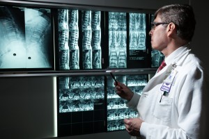
IMAGING TECHNIQUES FOR PAIN PROCEDURES
CT SCAN VS. FLUROSCOPY
Radiological imaging improves the safety and accuracy of injections and lesions in and around the spine to such an extent that radiological guided imaging is the standard of care.
Generally, I utilize a CT scanner for most procedures, maximizing safety, lesion accuracy, successful outcome, and length of outcome. CT is vastly superior to the conventionally used fluoroscopy for all the reasons above. The enhanced ability to visualize nerves, blood vessels, the spinal cord, cysts, facets, muscles, lungs, and the abdominal contents (combined with my vast experience and knowledge of CT guided procedures) has allowed me to perform safe and successful injections and lesions which few pain specialists would dream of attempting. These include lesions of the ganglia or “computers” of the spinal nerves—especially difficult at the top of the neck and in the thoracic spine (where the ribs are) —with excellent result in significantly helping patients with various painful disorders (certain headaches, neck pains, groin pains, pain following spinal fusions, and certain sacroiliac joint pains.) Other ganglia which are occasional targets for blocks and lesions are those controlling sympathetic nervous system activity, implicated in the treatment of some cases of RSD, or Complex Regional Pain Syndrome, as it is now called. These lie outside the spine—in front of the spine in the neck and to the side and front of the thoracic and lumbar spine—behind the lungs, kidneys, and bowels, respectively. Blocking or later radio-frequency lesioning the sympathetic ganglia in the neck and upper thoracic spine may help certain cases of this syndrome with arm pain. The same injections or lesions applied to the lumbar ganglia may help reduce leg pain in selected patients with Complex Regional Pain Syndrome affecting the leg. Drainage and decompression of nerves squeezed by synovial cysts is not possible without the help of CT or open surgery (Click here to go to “Surgery” section.) Use of CT scanning makes injection of Botox into the iliopsoas muscle—to the side of the lumbar spine and behind the bowels—safer and more accurate. Botox injections of this muscle may relieve certain muscle pain and cannot be performed safely in an office. Even with fluoroscopy, the risks of puncturing the bowel exists. For this reason this muscle is usually never injected with Botox, despite the fact that, in the right hands and in the appropriate patient, these injections may be quite helpful.
Visit us on... ![]()
![]()
Emile M. Hiesiger, M.D.
| The Corinthian |
| 345 East 37th Street |
| Suite 320 |
| New York, NY 10016 |
| • Phone (212) 697-1411 or |
| • Phone (212) 263-6123 |
| • Fax (212) 697-1399 |
Add us to your contacts. Snap below!

*instructions for qrCode use


